


杨晓虎医生的科普号
- 精选 孩子4岁总是咬下嘴唇,笑的时候门牙突还露牙龈,怎么办?
正常情况下,口腔周围有很多肌肉,如唇肌、舌肌及颊肌等,这些肌肉的相互平衡维持牙弓形态的正常。咬下唇习惯不良习惯,打破了口腔内外的肌肉平衡,上前牙总是咬在下唇的外侧,上前牙会对下唇及下颌前牙区产生向舌侧的力量,从而导致下前牙的舌倾、拥挤、下颌后缩,同时增加了对上前牙腭侧的压力,使上前牙向唇侧倾斜移位,出现牙间隙,上颌骨前部前突。在上、下前牙之间形成深覆盖,深覆盖,上下颌前牙咬合时看不到下牙。外观表现为上唇短而厚、上前牙前突和下颌后缩严重时上前牙覆盖在下唇外侧,导致正常状态下上下唇不能闭合,上前牙露在唇外,形成“开唇露齿”的错颌畸形状态。不及时治疗这种状况会持续加重。发现孩子有咬下唇的习惯,家长首先不能急躁,因为咬下唇的孩子有些表现有胆小、认生、内向,应首先采取心理治疗的方法,不要过多的指责孩子,多带孩子多参加集体、户外的活动,培养兴趣爱好,帮助孩子逐渐克服不良习惯。如果孩子的不良习惯很顽固,出现明显的牙颌发育异常,需要及时就诊,进行正畸治疗,这个孩子采用了肌功能矫正器,疗程6个月,获得很好的效果。本人在此郑重声明:不会在任何网站销售矫正器,也请大家不要相信网上售卖,要家长自行戴用的矫正器。治疗前:治疗后: 本文系朱红医生授权好大夫在线(www.haodf.com)发布,未经授权请勿转载。
朱红 主任医师 北京儿童医院 口腔科1.2万人已读 - 精选 唇腭裂患儿的喂养方法
方法1:注意体位:(1)取坐位或45゜角抱位,切忌平躺,以免呛咳;(2)采用面对面喂哺方式,以利观察。(3)采用俯卧位,使鼻腔在口腔上方而不致发生呛咳。方法2:孩子吸奶时,用手指堵住唇裂部位,帮助唇部闭合。方法3:选用十字开口的树胶奶瓶,因为十字型的开口在受到压迫时才会打开,孩子不会被呛到。方法4:采用挤喂的方式,即购买可以挤压的奶瓶或注射器,滴管喂养。方法5:通过吹气球,吸吮奶嘴或按摩肌肉训练颊,舌的功能。方法6:将奶嘴置于非裂开处,以免局部刺激过大。方法7:早期正畸治疗,如配戴一种由软硬两种树脂材料构成的Hotz矫治器,覆盖全部牙槽嵴和硬软腭,使口腔内形成负压并改善舌的运动,对喂哺有显著的改善。
丁桂聪 主任医师 深圳市儿童医院 口腔科2715人已读 - 精选 歪嘴哭常见问答
1.歪嘴哭是怎么一回事? 最近在网上遇到一些家长,他们的孩子有一种奇怪的问题:平静时看着好好的脸,一哭一笑却非常不自然。仔细观察,原来这些孩子在哭或者笑或者张大嘴时,一边的嘴角是歪的。 这就是俗称的歪嘴哭了。 歪嘴哭面容(Asymmetric crying facies,ACF)是指新生儿或婴儿的脸在哭闹时出现健侧嘴角下拉而患侧嘴角不动所产生的表情不对称。这种情况在新生儿中的发病率在3‰~8‰之间,平均大约160个新生儿中有1个。男女比例为0.83:1 ,也就是女性更多见。 2.歪嘴哭的病因是什么? 对歪嘴哭面容病因的认识一开始就认为是面瘫的一种。后来发现歪嘴哭面容患儿有相当一部分伴有心脏畸形,故而有人称之为“歪嘴哭综合征”。现在认为一小部分患儿和产伤有关,比如胎位不正或者胎儿过大造成生产时面颈部压力过大,损伤面神经。相反,早产儿和低体重儿发病率就低;多数患儿是发育畸形——特别是降口角肌和/或面神经下颌缘支的发育不全。 3. 孩子除了歪嘴哭还会有其他什么问题? 歪嘴哭面容的孩子合并心脏畸形、面颈部其他畸形(比如半侧颜面短小)的风险是普通孩子的3.5倍。所以,医生面诊是必要的,心脏彩超检查也是必要的。医生根据情况还可能要进一步做其他检查。 4. 歪嘴哭等孩子长大些会不会自己好? 如果是产伤,神经受压导致的歪嘴哭面容,是可以自己慢慢好转的,一般满月后就会开始缓解。大多数患儿是属于发育畸形,是不能自己好的。不过,随着年龄增长,周围肌肉可能有一部代偿功能,看起来会没有以前那么明显,只是大哭、大笑、大张口等剧烈表情的时候才比较明显。 5. 按摩、针灸等理疗方法有效吗? 理论上无效。出现效果的患儿一般还是周围肌肉的代偿功能。出现代偿的患儿,家长会以为按摩有效果,但这种代偿很多患儿都没有,所以这些家长无论多努力按摩都没有效果。 6. 孩子除了歪嘴哭,还有闭眼不紧、一边没有抬头纹、法令纹不对称、大小脸、一边耳朵畸形等等问题,是同样的病吗? 如果同时有上述的的部分或全部问题,就可能不是歪嘴哭面容,而是面瘫(面神经瘫痪)了。歪嘴哭面容可以单独出现,也可以是某些综合征的一部分。比如半侧颜面短小(又称第一二鳃弓综合征),也是我这里较常见的先天畸形,可以影响面神经总干,造成一侧面部瘫痪(包括嘴角),甚至影响颌骨发育、眼球发育。 7. 歪嘴哭可以治疗吗?怎么治? 歪嘴哭面容可以通过整形手术的方法进行治疗。目前主要分为以下方案:筋膜悬吊、神经转移和健侧麻痹。筋膜悬吊是从身体其他地方获取筋膜(比如大腿),通过移植一块双向的(平行的和垂直的)筋膜,使倾斜不对称的下唇恢复正常状态。优点是手术不复杂,创伤相对小;缺点是口角不能主动运动,是被下巴带动的。健侧麻痹要么切断健侧好的神经,要么用肉毒素之类麻痹好的神经,总之,是让好的这边也麻痹达到平衡的效果。好处是手术简单,创伤小。这几种方案各有利弊,病人或家长应当与医生详尽咨询,选择适合自己的治疗方案。 8. 什么时候治疗比较好? 治疗时间至少应等到患儿满月后,确定不是生产过程形成的神经压迫,才考虑手术治疗。满月以后,早期治疗效果更好,不主张等到6岁和其他方面整形手术一起进行。但若有严重的先天性心脏病,则先治疗先心为好。
陈亦阳 主任医师 广州市妇女儿童医疗中心 口腔颌面外科1.9万人已读 - 精选 粘液腺囊肿临床诊断及治疗
粘液腺囊肿是最常见的小唾液腺瘤样病变,好发于下唇及舌尖腹侧。 其病因常因为舌体运动受下前牙摩擦以及自觉或不自觉的咬下唇动作使粘膜下腺体受伤。临床表现为:囊肿位于粘膜下,表面仅覆盖一薄层粘膜,故呈半透明、浅蓝色的小泡。囊肿很容易被咬伤而破裂,流出蛋清样透明粘稠液体,囊肿消失。破裂处愈合后,又被粘液充满,再次形成囊肿。反复破损后不再有囊肿的临床特点,而表现为较厚的白色瘢痕状突起,囊肿透明度减低。治疗方式:目前最常用的治疗方式为手术切除。
项立 医师 重庆医科大学附属儿童医院 口腔科1万人已读 - 精选 唇腭裂的母乳喂养
一,喂养前的准备,平时应注意营养物质的吸收,有利于产生母乳,喂养前应从胸部开始做小圆圈的按摩,逐渐移向乳房周围以及乳头,以刺激更多的乳汁流向乳头。二,喂养过程,喂养时,应把婴儿鞋报纸与地面呈35度至45度角,用手指轻压乳晕,以使乳头翘起,易于被婴儿含住,在喂养过程中应相隔4-5分钟与一拍呗,以有助于吞咽于胃内的空气排出。因为母乳喂养,一般每次在12分钟以后才能够达到有效的吸吮时间,所以母乳喂养的时间,较人工喂养长,大约有45分钟左右。 本文系邓利琴医生授权好大夫在线(www.haodf.com)发布,未经授权请勿转载
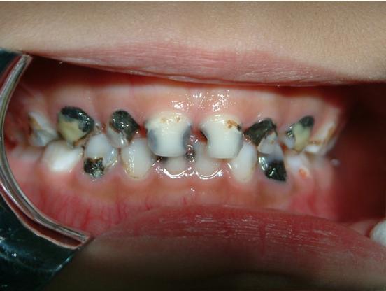
邓利琴 主任医师 广州市妇女儿童医疗中心 口腔科2262人已读 - 精选 乳牙为什么比恒牙更容易坏?
在工作中,牙医们常常会被问到这样的问题“这颗牙才长出来没多久,怎么就烂这么大个洞了?”,又或者是“这个牙怎么才长出来就是黑的?”、“小孩的牙齿是不是要比大人的软一点?”等等。这说明家长们也观察到了乳牙龋的一个重要特点:乳牙容易龋坏,并且龋坏进展速度非常快。事实上,乳牙一旦开始龋坏,从一个小黑点发展成大洞,仅仅需要三个月的时间。很多家长认为是小孩爱吃糖造成的,那么乳牙容易发生龋坏,且乳牙龋发展如此之快的真正原因又是什么呢? 其实乳牙本来就比恒牙更脆弱! 牙齿最表面的一层被称作牙釉质,是牙齿最坚硬的部分,就像牙齿的盔甲,抵御着外界各种的刺激。乳牙的牙釉质薄于恒牙,矿化的程度也更低,也就是说乳牙的保护壳厚度硬度都不够,难怪更容易坏了。 另外乳牙的形态也更容易使食物残渣滞留! 乳牙咬合面窝沟点隙多而深,牙齿之间随着颌骨生长又会出现缝隙,食物特别容易残留,稍不注意就刷不干净,成为细菌的温床,并且一旦发生了龋坏,牙齿表面变得粗糙了,就更不容易刷干净了。 再者儿童的饮食多为粘稠性强,含糖量高的软质食品,易发酵产酸 除了糖,各种蛋糕、面包、饼干等等也是容易使牙齿发生龋坏的食物哦。比起一次性吃很多甜食,孩子更喜欢一会吃一点,一会再吃一点,这样无意中延长了口腔细菌产酸破坏牙齿的时间。 最后儿童睡眠时间长,口腔处于静止状态,唾液分泌少,有利于细菌繁殖 唾液有促进牙釉质矿化的作用,也就是可以让牙齿的盔甲更坚硬,抵抗外界腐蚀,并且有清洁牙齿表面的作用。但是当我们睡觉的时候,唾液的分泌量会显著减少,同时细菌大量繁殖产酸,龋坏不知不觉就发生了。 本文系孙琪殷医生授权好大夫在线(www.haodf.com)发布,未经授权请勿转载
 孙琪殷 副主任医师 广州市妇女儿童医疗中心 口腔科1万人已读
 - 引用 摔伤后 如何预防疤痕
很多病人外伤后,急急忙忙跑到医院来,一来问我最多的两个问题,第一,这个伤口需不需缝针,第二就是会不会留疤。现代社会,对美观的要求越来越高。对于颌面部的损伤,如果处理不当,往往会造成疤痕增生,影响美观。我在这里给大家科普一下,我们家属如何来判断一个外伤需不需要缝针。首先看伤口有没有裂开,如果只是伤及皮肤表皮未伤及真皮层,粘膜未伤及粘膜下层,这种是不需要缝针的。若是伤及了皮下及粘膜下、肌肉层,但是伤口贴合得很紧,未暴露出口子,这样家属也可以考虑不缝针。换而言之,只要伤口裂开了,产生了口子,我们都建议缝针,或粘胶水,因为你伤口有多大,皮肤有多少不连续,将来就会长出多少疤痕来。那我们家属该如何来减少患儿的疤痕呢。1,伤口必须保持干燥、清洁,很多小朋友一来医院,一看伤口傻眼了,满伤口的药,全是碎渣,家属说自己涂点止血药。这无疑给清创带来了难度,增加伤口感染的机会,增加术后疤痕可能。这里必须给大家科普一下,如果出血,最好的方法是局部压迫。2,术中良好的复位,充分的减少张力,细针细线的缝合,皮内缝合技术的应用。3,术后伤口的护理同样极为重要。减张胶布的应用,这样可以控制疤痕。术后伤口的干洁,切勿湿水愈合之前,不要剧烈运动,出汗较多,污染伤口。待缝线脱落后局部的按摩,祛疤痕药物的使用。必要时皮下注射药物预防疤痕,后期激光美容。
罗冬元 主治医师 广州市妇女儿童医疗中心 口腔科1.1万人已读 - 引用 几岁孩子可以独立刷牙
养成良好的口腔卫生习惯要从娃娃抓起。 而在专家看来,许多父母在这方面做得并不到位。 许多父母认为孩子年纪太小,不敢让还在蹒跚学步的孩子自己洗澡,却敢让他们自己刷牙。 其实,由于孩子手部精细运动还有待发展,加上缺乏正确的引导,是很难自己刷干净牙齿的。 许多孩子成天乱吃零食、乱喝饮料,这些坏习惯会造成严重的牙齿龋坏。你知道吗?预防蛀牙和其它口腔问题,越早越好。 专家给父母们的建议如下: 每次给宝宝喂完食物,都要记得用柔软的纱布清洁宝宝的牙龈;不要在奶瓶里灌上奶或果汁,然后让孩子含着奶瓶睡觉。一定不要因为孩子喜欢,就把奶嘴蘸上蜂蜜或者别的糖浆给孩子含着。 致龋菌感染性强,可以很容易地通过勺子或奶嘴传给孩子,所以,不要让老人或保姆共用宝宝的餐具,或者亲吻宝宝的嘴唇。 不要让孩子整天乱吃零食——即使是最健康的零食,也会在嘴里留下足以喂饱致龋菌的食物残渣。如果要给孩子吃零食,也应该选择水果或是其它未经加工的食物,而不是袋装垃圾食品,以及橡皮糖、果丹皮和果干之类粘牙的东西。 果汁(即使是有机果汁),因为含糖量非常高,对于牙齿来说也和碳酸饮料一样是一种危险的饮料。尽量用水代替。 3岁前,使用米粒大小的含氟牙膏给孩子刷牙,一天两次。3岁后可以改为豌豆大小的牙膏。 孩子应该从几岁开始自己完全独立刷牙,因人而异。2岁可以开始训练,等到孩子会自己系鞋带或者梳马尾辫了再说吧(一般7岁以后)。 同时,需要使用菌斑检知方法确认孩子其实可以独立刷干净了才可以放手让孩子自己操作。即使自己开始操作,也需要家长每天确认是不是刷干净了,不能放羊。 7岁前的刷牙应该是孩子自己刷一遍,然后家长再给孩子认认真真刷一遍。 在孩子长出两颗相邻的牙齿以后,就应该开始使用牙线清洁牙缝了。 本文系李爽医生授权好大夫在线(www.haodf.com)发布,未经授权请勿转载 。
李爽 主治医师 北京儿童医院 口腔科8335人已读 - 引用 全口涂氟后注意事项
6岁以下儿童推荐选择2.26%含氟浓度的氟保护漆◇全口涂氟后4小时后才可进食固体食物;◇涂氟当天不要刷牙、漱口;◇涂氟不能替代日常使用含氟牙膏刷牙;根据个人患龋风险,需要每3-6个月定期涂氟以维持疗效;本文系张增方医生授权好大夫在线(www.haodf.com)发布,未经授权请勿转载
1.9万人已读 - 引用 国际牙外伤协会的年轻恒牙外伤管理指南1(2007年版)(张增方译制初稿完善中)
Guidelines for the managementof traumatic dental injuries. I. Fractures and luxations of permanent teeth牙外伤指南1 恒牙折断及脱位Fig 1. (a) A 9-year-old girl visiting the emergency dental clinic 30 min after falling from a bicycle. (b) (close up view of Fig. 1a) Clinical examination showing lateral luxation of the left central incisor with fracture of the alveolar process. The incisor is luxated to a superior and labial position.图1(a)9岁女孩儿从单车上摔下,30分钟后到急诊科就诊。临床检查可见左上中切牙侧方脱位,伴有牙槽突骨折。中切牙向上唇侧方向脱位。Fig. 2. Crown fracture of rightcentral incisor and crown-root fracture of left central incisor.图2 右侧上中切牙冠折及左侧上中切牙冠根折Fig. 3. Intrusive luxation ofright lateral and central incisors. Crown fractures are seen on both intrudedincisors and theadjacent left central incisor.图3右侧侧切牙及中切牙挫入性脱位。两个挫入牙及左侧中切牙见冠折。一、年轻恒牙牙折及牙槽突骨折的治疗指南Treatment guidelines forfractures of teeth and alveolar bone Clinical findings Radiographic findings Treatment Uncomplicated crown fracture Fracture involves enamel or dentin and enamel; the pulp is not exposed. Sensibility testing may be negative initially indicating transient pulpal damage; monitor pulpal response until a definitive pulpal diagnosis can be made 非复杂性冠折 牙折累及釉质或釉本质;未露髓。 敏感试验最初可能阴性,意味着暂时性的牙髓创伤,需监控牙髓反应直到诊断明确 The 3 angulations described in radiographic examination to rule out displacement or fracture of the root. Radiograph of lip or cheek lacerations is recommended to search for tooth fragments or foreign material 三角度牙片??来明确牙移位及根折情况。 建议唇颊部软组织影像来排除牙碎片及异物 If tooth fragment is available, it can be bonded to the tooth. Urgent care option is to cover the exposed dentin with a material such as glass ionomer or a permanent restoration using a bonding agent and composite resin. Definitive treatment for the fractured crown may be restoration with accepted dental restorative materials 如果能找到冠折片可将其粘结于牙齿。 紧急处理措施是尽快覆盖暴露的牙本质,可用玻璃或粘结剂加永久树脂恢复冠外形。 明确的治疗方案是:用可接受的材料恢复冠外形。 Complicated crown fracture Fracture involves enamel and dentin and the pulp is exposed. Sensibility testing is usually not indicated initially since vitality of the pulp can be visualized. Follow-up control visits after initial treatment includes sensibility testing to monitor pulpal status 复杂性冠折 牙折累及釉质或釉本质;已露髓。 ??最初的敏感试验常常 不明确因为可见活髓。 初诊治疗结束后应多次复诊进行明显敏感试验仪明确牙髓状态 The 3 angulations described in radiographic examination to rule out displacement or fracture of the root. Radiograph of lip or cheek lacerations is recommended to search for tooth fragments or foreign material. The stage of root development can be determined from the radiographs 三角度牙片??来明确牙移位及根折情况。 建议唇颊部软组织影像来排除牙碎片及异物 影像学可明确牙根的发育阶段 In young patients with immature, still developing teeth, it is advantageous to preserve pulp vitality by pulp capping or partial pulpotomy. This treatment is also the choice in young patients with completely formed teeth. Calcium hydroxide and MTA (white) are suitable materials for such procedures. In older patients, root canal treatment can be the treatment of choice, although pulp capping or partial pulpotomy may also be selected. If too much time elapses between accident and treatment and the pulp becomes necrotic, root canal treatment is indicated to preserve the tooth. In extensive crown fractures a decision must be made whether treatment other than extraction is feasible 对于年轻恒牙应尽量通过盖髓术或冠髓切断术来保存活髓; 对于牙根发育完全的年轻患者也可采取通过盖髓术或冠髓切断术来保存活髓; 氢氧化钙或MTA是合适的材料; 对于年龄稍大的患者可考虑选择RCT,然而仍可采用盖髓术或冠髓切断术。 如果外伤就诊时间太迟已经发生牙髓坏死, RCT是明确的治疗方案。 对于大面积的冠折,应考虑是否治疗还是拔除。? Crown-root fracture Fracture involves enamel, dentin and root structure; the pulp may or may not be exposed. Additional findings may include loose, but still attached, segments of the tooth (Fig. 2). Sensibility testing is usually positive 冠根折 牙折累及釉质,牙本质及根部;可能露髓或未露髓;检查可及松动,但折断片仍与牙连接(图2) 敏感试验阳性 As in root fractures, more than one radiographic angle may be necessary to detect fracture lines in the root (see radiographic examination) 必要时可采用多角度牙片判断根折线的位置 Treatment recommendations are the same as for complicated crown fractures (see above). In addition, attempts at stabilizing loose segments of the tooth by bonding may be advantageous, at least as a temporary measure, until a definitive treatment plan can be formulated 治疗建议同复杂性冠折 另外,可尝试固定粘结折断片,只少可做为在明确治疗方案之前的一种临时性的方法 《牙外伤手册》26页中B建议切除腭侧牙槽骨,是否值得这么做,种植医师的意见?? 冠根折的治疗目的多是为后期做桩核冠修复而延长根使其达龈上做准备 Root fracture The coronal segment may be mobile and may be displaced. The tooth may be tender to percussion. Sensibility testing may give negative results initially, indicating transient or permanent pulpal damage; monitoring the status of the pulp is recommended. Transient crown discoloration (red or grey) may occur 根折 冠断片可能松动或移位,叩痛明显,最初敏感试验阴性,意味着牙髓创伤,建议监测牙髓状态,可能出现暂时性的牙冠变色 The fracture involves the root of the tooth and is in a horizontal or diagonal plane. Fractures that are in the horizontal plane can usually be detected in the regular 90_ angle film with the central beam through the tooth. This is usually the case with fractures in the cervical third of the root. If the plane of fracture is more diagonal, which is common with apical third fractures, an occlusal view is more likely to demonstrate the fracture including those located in the middle third 根部涉及水平吧或斜面的折断。水平折断常常可由90度投照可见,此种折断常见于颈部根折;呈斜面的折断在根中或根尖部折断常见,可采用咬合片协助诊断 Reposition, if displaced, the coronal segment of the tooth as soon as possible. Check position radiographically. Stabilize the tooth with a flexible splint for 4 weeks. If the root fracture is near the cervical area of the tooth, stabilization is beneficial for a longer period of time (up to 4 months). It is advisable to monitor healing for at least 1 year to determine pulpal status. If pulp necrosis develops, root canal treatment of the coronal tooth segment to the fracture line is indicated to preserve the tooth 若存在移位,尽可能将冠方部分复位,并拍定位牙片,将患牙弹性固位4周。 若根折靠近牙颈部,应将固定时间延长(可4个月以上)。 复查牙髓状态1年,若出现牙髓坏死,行冠方部分RCT以保留患牙。 ??为什么只做冠方部分,若颈部及中不折断也是这样吗??根部髓不会感染吗??不会残髓炎吗?临床具体怎么操作?才知道刚刚好做到了根折处。 Alveolar bone fracture The fracture involves the alveolar bone and may extend to adjacent bone. Segment mobility and dislocation are common findings. An occlusal change due to misalignment of the fractured alveolar segment is often noted. Sensibility testing may or may not be Positive 牙槽突骨折 骨折累及牙槽骨,且可累及临骨?? 常存在折断片松动及移位,咬合关系改变, 牙敏感试验阴性或阳性 Fractures lines may be located at any level, from the marginal bone to the root apex. The panoramic technique is of great help in determining the course and position of fracture lines 骨折线可能位于从骨边缘(牙颈部)到根尖的任何水平。 全景片可明确骨折线的位置及走向。 Reposition any displaced segment and then splint. Stabilize the segment for 4 weeks 移位断片复位及夹板固定。 固定4周后拆除。 恒牙折断及牙槽突折断的复查程序 Time 4 weeks 6–8 weeks 4 months 6 months 1 year 5 years Uncomplicated crown fracture C(1) C(1) Complicated crown fracture C(1) C(1) Crown-root fracture C(1) C(1) Root fracture S + C(2) C(2) S(*) + C(2) C(2) C(2) C(2) Alveolar fracture S + C(3) C(3) C(3) C(3) C(3) C(3) S, splint removal.S (*), splint removal incervical third fractures.C, clinical and radiographicexamination恒牙折断及牙槽突折断一些好的或坏的预后情况 Favorable outcome Unfavorable outcome 1 Asymptomatic; positive response to pulp testing; continuing root development in immature teeth. Continue to next evaluation 无症状;活力测阳性; 年轻恒牙牙根继续发育; 继续评估; Symptomatic; negative response to pulp testing; signs of apical periodontitis; no continuing root development in immature teeth. Root canal treatment is indicated 有临床症状;活力测阴性;有根尖周炎的迹象;年轻恒牙牙根未能继续发育;需RCT 2 Positive response to pulp testing (false negative possible up to 3 months). Signs of repair between fractured segments. Continue to next evaluation Negative response to pulp testing (false negative possible up to 3 months). Clinical signs of periodontitis. Radiolucency adjacent to fracture line. Root canal treatment is indicated only to the line of fracture(临床上怎么操作达到这个?) 活力测阴性(可能3月以上假阴性??),有根尖周炎的临床症状;邻近折断线处见低密度影。RCT只达折断处,保留根髓 3 Positive response to pulp testing (false negative possible up to 3 months). No signs of apical periodontitis. Continue to next evaluation 活力测阳性(3月以上假阴性); 无根尖周炎的signs迹象 继观评估 Negative response to pulp testing (false negative possible up to 3 months). Signs of apical periodontitis or external inflammatory resorption. Root canal treatment is indicated 活力测阴性(可能3月以上假阴性); 有根尖周炎及根外表吸收的迹象;(见《牙外伤手册》51页关于“感染引起根吸收的治疗”) 行RCT。 二、牙脱位治疗指南Treatment guidelines for luxation injuries Clinical findings Radiographic findings Treatment Concussion The tooth is tender to touch or tapping; it has not been displaced and does not have increased mobility. Sensibility tests are likely to give positive results 牙震荡 牙齿轻触敏感,叩痛明显,无移位及松动。敏感试验阳性。 No radiographic abnormalities 无影像学异常 No treatment is needed. Monitor pulpal condition for at least 1 year 无需处理。复查牙髓状况至少一年。 Subluxation The tooth is tender to touch or tapping and has increased mobility; it has not been displaced. Bleeding from gingival crevice may be noted. Sensibility testing may be negative initially indicating transient pulpal damage. Monitor pulpal response until a definitive pulpal diagnosis can be made 亚脱位(牙松动) 牙齿轻触敏感,叩痛明显;有一定松动但无移位; 可见龈沟出血; 敏感试验最初可阴性,表示暂时性牙髓创伤; 需复查牙髓反应直到明确牙髓诊断 Radiographic abnormalities are usually not found 影像学常无异常。 A flexible splint to stabilize the tooth for patient comfort can be used for up to 2 weeks 采用弹性夹板固定至少2周,以减轻患者不适。 Extrusive luxation The tooth appears elongated and is excessively mobile. Sensibility tests will likely give negative results. In mature teeth, pulp revascularization some times occurs. In immature, not fully developed teeth, pulpal revascularization usually occurs 牙部分脱位 牙伸长,松动明显。 敏感试验可能阴性。 在发育完成的恒牙有时也可出现牙髓血管再生;在年轻恒牙常可出现牙髓再生。 Increased periodontal ligament space apically 根尖部可见牙周膜间隙增宽 Reposition the tooth by gently re-inserting it into the tooth socket. Stabilize the tooth for 2 weeks using a flexible splint. Monitoring the pulpal condition is essential to diagnose root resorption. In immature developing teeth, revascularization can be confirmed radiographically by evidence of continued root formation and pulp canal obliteration and usually return to response to sensibility testing. In fully formed teeth, a continued lack of response to sensibility testing should be taken as evidence of pulp necrosis together with periapical rarification and sometimes crown discoloration 用轻柔力将脱位牙复位于牙槽窝;弹性夹板固位脱位牙2周; ??定期复查牙髓状态对诊断根吸收是必要的(?牙髓坏死感染是造成根吸收的原因)(恒牙出现根吸收的机会不大吧?预后一般是:年轻恒牙出现根管闭锁,成熟恒牙出现牙髓坏死,且年轻恒牙预后要好些) 对于年轻恒牙可通过X线片复查确定是否出现牙髓血管再生,如果有再生则牙根会继续发育及出现根管闭锁,而且常常会出现敏感试验恢复正常。 对于成熟恒牙,持续的敏感试验无反应就意味着牙髓坏死,可能伴随出现根尖周rarification??,有时还会出现牙冠变色。 Lateral luxation The tooth is displaced, usually in a palatal/lingual or labial direction (Fig. 1a, b). It will be immobile and percussion usually gives a high, metallic (ankylotic) sound. Sensibility tests will likely give negative results. In immature, not fully developed teeth, pulpal revascularization usually occurs 侧方移位 牙齿出现了向唇/腭向或侧方移位;(视) 牙齿不松,叩诊呈高调固连音;(触叩) 敏感试验阴性; 年轻恒牙常出现牙髓再生 The widened periodontal ligament space is best seen on eccentric or occlusal exposures 咬合片可见牙周膜间隙增宽 Reposition the tooth with forceps to disengage it from its bony lock and gently reposition it into its original location. Stabilize the tooth for 4 weeks using a flexible splint. Monitor the pulpal condition. If the pulp becomes necrotic, root canal treatment is indicated to prevent root resorption. In immature, developing teeth, revascularization can be confirmed radiographically by evidence of continued root formation and possibly by positive sensibility testing. In fully formed teeth, a continued lack of response to sensibility testing indicates pulp necrosis, along with periapical rarification and sometimes crown discoloration 牙钳松解骨内锁结,予轻柔复位到原来的位置; 弹性夹板固定4周; 复查牙髓状态;如果出现牙髓坏死,及时行RCT以避免牙根吸收??(牙髓坏死与牙根吸收的关系,成熟恒牙也容易出现牙根吸收??临床见到的多吗?) 对于年轻恒牙可通过X线片检查牙根继续发育的情况来确定是否出现牙髓血管再生,也可通过牙髓反应来证明。 对于成熟恒牙,持续的敏感试验无反应就意味着牙髓坏死,可能伴随出现根尖周rarification??,有时还会出现牙冠变色。 Intrusive luxation The tooth is displaced axially into the alveolar bone. It is immobile and percussion may give a high, metallic (ankylotic) sound (Fig. 3). Sensibility tests will likely give negative results. In immature, not fully developed teeth, pulpal revascularization may occur 牙挫入 牙齿轴向移位至牙槽骨; 牙齿不松,叩诊呈高调固连音; 敏感试验阴性; 年轻恒牙可能出现牙髓再生 The periodontal ligament space may be absent from all or part of the root 牙周膜间隙完全消失或部分消失 1. Teeth with incomplete root formation: Allow spontaneous repositioning to take place. If no movement is noted within 3 weeks, recommend rapid orthodontic repositioning. 2. Teeth with complete root formation: The tooth should be repositioned either orthodontically or surgically as soon as possible. The pulp will likely be necrotic and root canal treatment using a temporary filling with calcium hydroxide is recommended to retain the tooth 1. 年轻恒牙:可任其自行萌出;若3周内无任何移出,建议快速正畸牵出。 (局麻下轻轻摇松解除锁结,任其自行萌出,,或正畸牵引,保证3周内复位,这样一旦发生牙髓坏死或炎性吸收可及时RCT----- 40页 《牙外伤》葛立宏译) 2. 成熟恒牙:应尽快正畸复位或外科手术复位;一般都会发生牙髓坏死,应行RCT,并建议暂时用氢氧化钙充填根管以保留患牙。 (不可能自行萌出;可外科复位或正畸牵引复位;正畸牵引应在3周内完成;之后行预防性牙髓摘除;需密切观察牙折牙髓并发症----40页 《牙外伤》葛立宏译) Avulsion 牙完全脱出属于另一种不同的系列 见年轻恒牙外伤指南2 Follow-up procedures forluxated permanent teeth脱位恒牙的复查程序 Time 至少2w 4w 6-8w 6m 1y 5y内每年复查 牙震荡/亚脱位 C(1) C(1) C(1) NA 牙部分脱出 S+C(2) C(3) C(3) C(3) C(3) C(3) 牙侧方移位 C(3) S C(3) C(3) C(3) C(3) 牙挫入 C(4) C(4) C(4) C(4) C(4) S, splint removal.夹板拆除 C, clinical andradiographic examination.临床及X线检查 NA, not applicable.Favorable and unfavorableoutcomes include some, but not necessarily all of the following良性和不良预后 Favorable outcomes unfavorable outcomes 1 Asymptomatic; positive response to pulp testing (false negative possible up to 3 months); continuing root development in immature teeth; intact lamina dura?? 无临床症状; 牙髓活力测阳性(可能出现3个月以上的假阳性); 年轻恒牙的牙根继续形成; 牙周膜完好?完美的根尖周表现? Symptomatic; negative response to pulp testing (false negative possible up to 3 months); no continuing root development in immature teeth, periradicular radiolucencies 有临床症状; 牙髓活力测阴性(3个月以上的假阴性??); 年轻恒牙牙根停止发育; 根尖周出现低密度影; 2 Minimal symptoms; slight mobility; no excessive radiolucency periradicularly 轻微的临床症状; 轻度松动; 根尖周无低密度影 Severe symptoms; excessive mobility; clinical and radiographic signs of periodontitis. Root canal treatment is indicated in a closed apex tooth. In immature teeth, apexification procedures are indicated 严重的临床症状; 严重的松动; 临床及X线支持根尖周炎; 成熟恒牙行RCT; 年轻恒牙行根尖诱导术 3 Asymptomatic; clinical and radiographic signs of normal or healed periodontium; positive response to pulp testing (false negative possible up to 3 months). Marginal bone height corresponds to that seen radiographically after repositioning 无临床症状; 临床及X线检查正常,牙周愈合良好; 牙髓活力测试正常(3月以上的假阳性) 牙槽突边缘高度与刚复位时一致 Symptoms and radiographic sign consistent with periodontitis; negative response to pulp testing (false negative possible up to 3 months); breakdown of marginal bone??. Splint for additional 3- to 4- week period; root canal treatment is indicated if not previously initiated??; chlorhexidine mouth rinse 临床表现及X线显示根尖周炎; 牙髓活力测试阴性(3个月以上的假阳性); 牙槽突出现吸收, 继续夹板固定3到4周, 行RCT? 4 Tooth in place or erupting; intact lamina dura; no signs of resorption. In mature teeth, start the root canal treatment within the first 3 weeks 牙就位并再萌; 根尖周完好?,未出现牙根吸收; 成熟恒牙在3周内开始RCT Tooth locked in place/ankylotic tone??; radiographic signs of apical periodontitis; external inflammatory resorption or replacement resorption? 牙固连? X线诊断根尖周炎; 外缘炎性吸收或内吸收?? Splinting guidelines for tooth/bone fractures andluxated/avulsed teeth三、牙/骨折断及脱位/脱出的夹板固定指南Splinting times 固定时间Type of injury Splinting timeSubluxation 2 weeksExtrusive luxation 2 weeksAvulsion 2 weeksLateral luxation 4 weeksRoot fracture (middlethird) 4 weeksAlveolar fracture 4 weeksRoot fracture (cervicalthird) 4 monthsType of splints 固定的种类Acid-etch bonded compositesplints are recommended, e.g. wire-composite splints and TTS (titanium traumasplint). For detailed description of splinting see current textbooks andarticles推荐全酸树脂夹板固定,例如加强丝-树脂夹板或外科钛丝夹板。夹板的详细描述详见文献。References1.Andreasen JO, Andreasen F, Andersson L. Textbook and color atlas of traumaticinjuries to the teeth, 4th edn.Oxford:Blackwell Munksgaard; 2007.2.Petersson EE, Andersson L, Sorensen S. Traumatic oral vs non-oral injuries.Swed Dent J 1997;21:55–68.3. GlendorU, Halling A, Andersson L, Eilert-Petersson E. Incidence of traumatic toothinjuries in children and adolescents in the county of Vastmanland, Sweden. SwedDent J 1996;20:15–28.4.Flores MT, Andreasen JO, Bakland LK et al.. International Association of DentalTraumatology. Guidelines for the evaluation and management of traumatic dentalinjuries. Dent Traumatol 2001;17:1–4.5.Andreasen JO, Andreasen F, Bakland L, Flores MT. Traumatic dental injuries. Amanual, 2nd edn. Oxford: Blackwell Munksgaard; 2003.6.Andreasen JO, Andreasen FM, Skeie A, Hjorting-Hansen E, Schwartz O. Effect oftreatment delay upon pulp and periodontal healing of traumatic dental injuries- a review article. Dent Traumatol 2002;18:116–28.7.Andreasen JO, Andreasen FM, Mejare I, Cvek M. Healing of 400 intra-alveolarroot fractures. 1. Effect of pre-injury and injury factors such as sex, age,stage of root development, fracture type, location of fracture and severity of dislocation.Dent Traumatol 2004;20:192–202.8.Cvek M, Meja`re I, Andreasen JO. Conservative endodontic treatment of teethfractured in the middle or apical part of the root. Dent Traumatol2004;20:261–269.9.Cvek M, Andreasen JO, Borum MK. Healing of 208 intraalveolar root fractures inpatients aged 7–17 years. Dent Traumatol 2001;17:53–62.10.Jackson NG, Waterhouse PJ, Maguire A. Factors affecting treatment outcomesfollowing complicated crown fracturesmanagedin primary and secondary care. Dent Traumatol 2006;22:179–85.11.Rafter M. Apexification: a review. Dent Traumatol 2005;21:1–8. Review.12.Andreasen JO, Bakland LK, Andreasen FM. Traumatic intrusion of permanent teeth.Part 3. A clinical study of theeffectof treatment variables such as treatment delay, method of repositioning, typeof splint, length of splinting and antibiotics on 140 teeth. Dent Traumatol2006;22:99– 111.13.Andreasen JO, Bakland LK, Andreasen FM. Traumatic intrusion of permanent teeth.Part 2. A clinical study of the effect of preinjury and injury factors, such assex, age, stage of root development, tooth location, and extent of injury includingnumber of intruded teeth on 140 intruded permanent teeth. Dent Traumatol2006;22:90–8.14.Andreasen JO, Bakland LK, Matras RC, Andreasen FM. Traumatic intrusion ofpermanent teeth. Part 1. An epidemiological study of 216 intruded permanentteeth. Dent Traumatol 2006;22:83–9.15.Filippi A, von Arx T, Lussi A. Comfort and discomfort ofdental trauma splints -a comparison of a new device (TTS)withthree commonly used splinting techniques. Dent Traumatol 2002;18:275–80.16.Von Arx T, Filippi A, Lussi A. Comparison of a new dental trauma splint device(TTS) with three commonly usedsplintingtechniques. Dent Traumatol 2001;17:266–74.17.Von Arx T, Filippi A, Buser D. Splinting of traumatized teeth with a newdevice: TTS (Titanium Trauma Splint). Dent Traumatol 2001;17:180–4.
8935人已读

杨晓虎主治医师
徐州市儿童医院口腔科
